Research overview
We are carrying out some research In Cellular biochemistry laboratory (Nagahara Lab) about the apoptosis which is the main form of cell death.
Apoptosis is a phenomenon that eradicates the cells(for example, injured or dead cells that are no longer working properly) which are no longer useful for living organisms to maintain a healthy life (It is called “homeostasis”) . The beginning of apoptosis is not only occurred by the determination of own cells, but it’s also occurred by the dictate of other cells. As human beings, we are consisted of 60 trillion cells which are not living licentiously, but living coordinately, so that all human beings are able to survive. If apoptosis is no longer working properly, it may result in the increasing risks of illness. For example, when apoptosis becomes less likely to occur, the cells that should have been perished or have become strange by injuring will increase and become cancer cells. On the contrary, if apoptosis is liable to occur in brain, It has been known that such as Alzheimer's diseases will be caused. Just as the theory that anomalous of apoptosis is involved in about 60% of the human diseases, there is a deep relation between apoptosis and maintenance of our homeostasis.
Since there are therapeutic benefits to cure diseases by preventing the abnormality of apoptosis, we are studying how apoptosis is triggered and how the signal transduction occurs between the cells by using biochemistry, cell biology and molecular biology techniques.
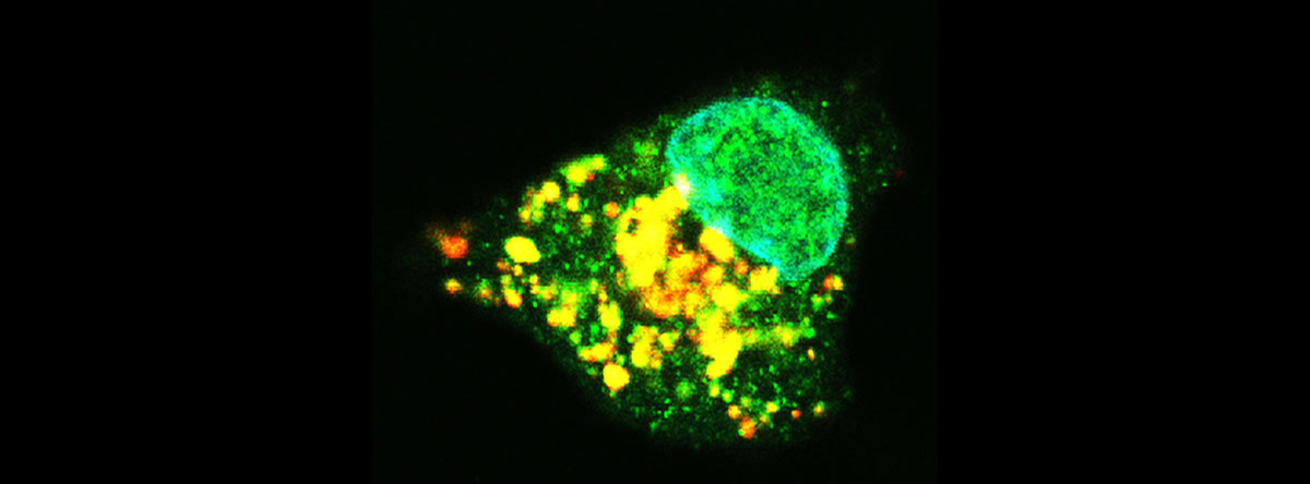
As one of the organelle presents in cell, mitochondria not only takes charge of the production of energy in cells, but also plays an important role in apoptosis. Moreover, it has been being apparent recently that Injured mitochondria was packaged by biomembrane and fusing with lysosome, which lead to intracellular phagocytosis and elimination of cells to cause mitophagy - one kind of autophagy that works for maintaining the cell survival. Hence, We can say that as an organelle, mitochondria is deeply related to cell survival and cell death.
Pyknosis and cell morphological changes along with the pyknosis is as a function of apoptosis. As a feature of apoptosis, there is the nuclear condensation and the associated cell morphological change. This is mainly due to the specifically activation of these groups of enzymes and caspase during apoptosis.
One of the reason for the activation of caspase is that the cytochrome c is released into cytoplasm, which is usually present in mitochondria, and related to the energy production. The released cytochrome c enables the caspase activation and the proceeding of apoptosis. So, what kind of substances release the cytochrome c from mitochondria ? So far, such as oxidative stress and special protein (Bcl-2 family proteins) which proceed the release of cytochrome c by binding to the mitochondria has became clear.
In addition, it has become apparent that Bcl-2 inhibits apoptosis and mitophagy . In the contrary, we can consider from the Bax and Beclin-1 inducement to apoptosis and mitophagy that Bcl-2 and Bcl-2 family proteins gather in mitochondria and then response the extent of injury to change-over switch of cell survival and cell death.
So far, we have shown that the lipids and similar substances present in cells induce the release of cytochrome c by acting on mitochondria. So we are working on to elucidate the signaling mechanism to mitochondria by lipid. Such compounds like anti-cancer agents and immunosuppressive agents, which could working on the elimination of certain cells by apoptosis, were expected to be applied. In addition, it has become the key on how to inhibit cancer cells from causing mitophagy. Therefore, by collaborating with other laboratories, we are also studying the development of drugs that induce apoptosis effectively and the elucidation of the mechanism of induction.
Related Papers: Apoptotic cells are eaten by special cells called phagocytes. If this kind of mechanism becomes aberration, it would remain apoptotic cells without being eaten, and would lead to the inflammation of the surrounding tissues. So that the eradication of no functional cells is very vital for health maintenance . We are studying about the improvement of phagocytosis of phagocytic cells and the mechanism of phagocytosis primarily in apoptotic cells.
Related Papers: Stratum corneum of skin consists of corneocyte (differentiated keratinocyte) and intercellular lipids that are filled between them . The purpose of the Stratum corneum is to form a barrier to protect underlying tissues from invasion of foreign substances and maintain the function of Water conservation.
On the other hand, it has become clear that there are 14 types of caspase that are activated during apoptosis. However, it has become apparent that caspase 14 is abundant in keratinocytes, and is related to maintain the barrier function and Water conservation function of the Stratum corneum by creating a natural moisturizing factor and decomposing the filaggrin precursor independent of apoptosis. In short, a compound that activates caspase 14 and induces its expression effectively, leads to the amelioration of skin function. Therefore, we are working on to clarify the skin differentiation mechanism by exploring the mechanism of the substance that causes the expression inducement of caspase 14.
Related Papers: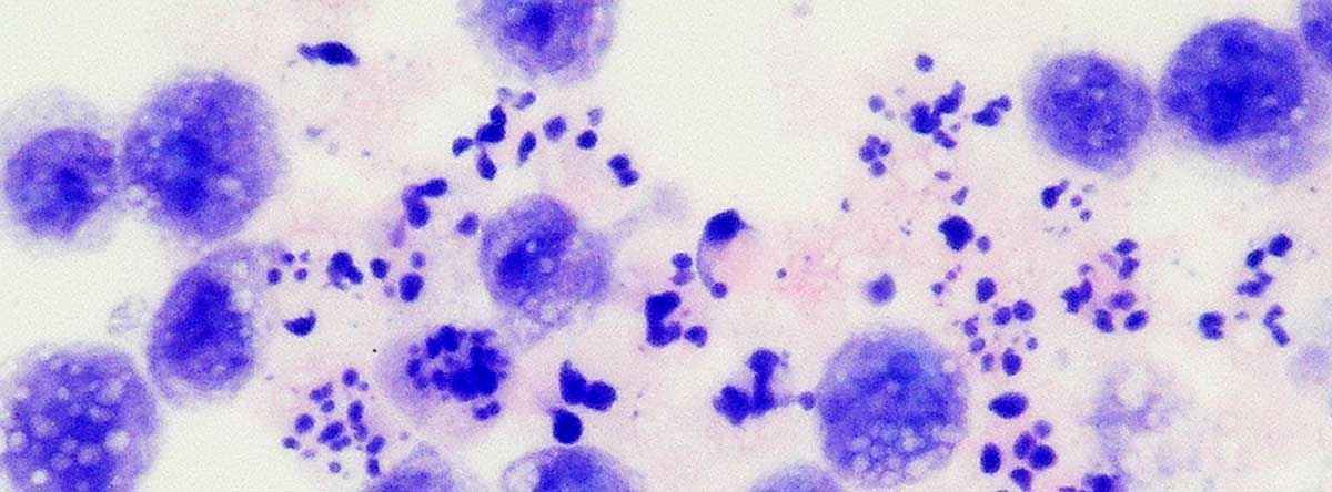
1. The mechanism of apoptosis and autophagy
・Nagahara Y., Takeyoshi M., Sakemoto S., Shiina I., Nakata K., Fujimori K., Wang Y., Umeda E., Watanabe C., Uetake S., Yamori T., Dan S., Yoshimi Y., Shinomiya T., Ikekita M. Novel tamoxifen derivative Ridaifen-B induces Bcl-2 independent autophagy without estrogen receptor involvement. Biochem. Biophys. Res. Comm., Vol.435, No.4, 657-663, 2013.
・Nagahara Y., Suzuki E., Sekine Y., Uchiro H., Yoshimi Y., Shinomiya T., Ikekita M. SUTAF, a novel β-methoxyacrylate derivative, promotes neurite outgrowth with extracellular signal-regulated kinase and c-jun N-terminal kinase activation. Eur. J. Pharmacol., Vol.694, No.1-3, 53-59, 2012.
・Fukui M., Nagahara Y., Nishio Y., Honjo T., Shinomiya T. Induction of Mitochondria Defect and Caspase-dependent Apoptosis to human T-cell leukemia Jurkat cells by Rokitamycin. J. Pharmaco. Sci., Vol.110, No.1, 69-77, 2009.
・Nagahara Y., Shiina I., Nakata K., Sasaki A., Miyamoto T., Ikekita M. Induction of Mitochondria-Involved Apoptosis in Estrogen Receptor Negative Cells by a Novel Tamoxifen Derivative, Ridaifen-B. Cancer Sci., Vol.99, No.3, 608-614, 2008.
・Nagahara Y., Tanaka M., Shinomiya T. Mechanism of Mitochondrial 7A6 Antigen Exposure Triggered by Distinct Apoptotic Pathways: Involvement of Caspases. Cytometry, Part A, Vol. 71A, No.4, 232-241, 2007.
・Nagahara Y., Shinomiya T., Kuroda S., Kaneko N., Nishio R., and Ikekita M. Phytosphingosine induced mitochondria-involved apoptosis. Cancer Sci. Vol. 96, No. 2, 83-92, 2005. 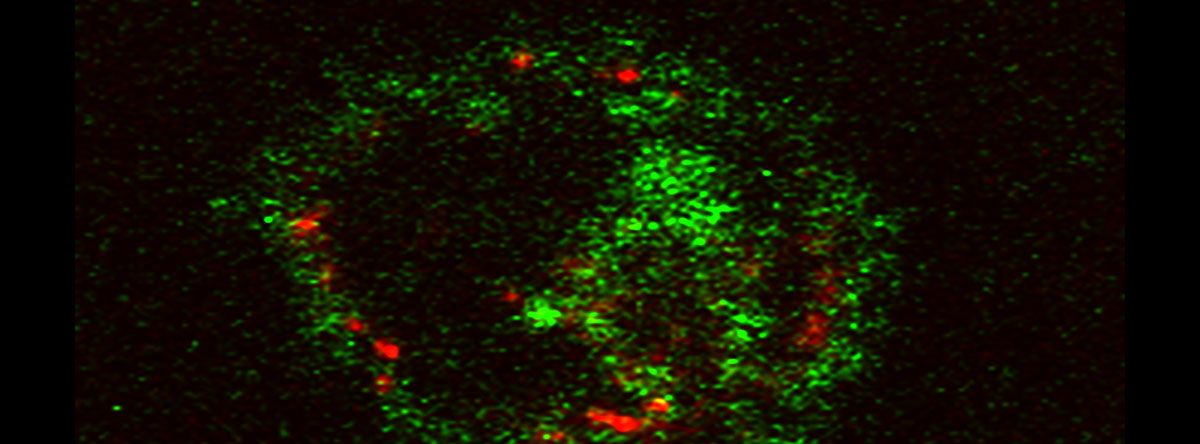
2. The mechanism of phagocytosis of apoptotic cells
・Tamegai H., Takada Y., Okabe M., Asada Y., Kusano K., Katagiri Y.U., Nagahara Y. Aureobasidium pullulans culture supernatant significantly stimulates R-848-activated phagocytosis of PMA-induced THP-1 macrophages. Immunopharm. Immunotoxicol., in press.
・Nagahara Y., Nagamori T., Tamegai H., Hitokuwada M., Yoshimi Y., Ikekita M., Shinomiya T. Inulin stimulates phagocytosis of PMA-treated THP-1 macrophages by involvement of PI3-kinases and MAP kinases. Biofactors, Vol.37, No.6, 447-454, 2011. 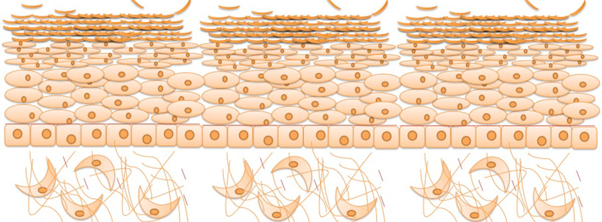
3. Elucidation of the mechanism of skin differentiation
・Nagahara Y., Kawakami K., Sikandan A., Yagi D., Nishikawa R., Shinomiya T. Sphingoid base-upregulated caspase-14 expression involves MAPK. Biol. Pharm. Bull.,Vol. 41, No. 5, 743-748, 2018.